
Coronary Angiography in Aurangabad
Opening Hour's
-
Monday - Saturday
9:00 AM To 9:00 PM -
Sunday
Closed
Quick Contact
Reach Us
Office No 3, 2nd floor, Mulay-Tapadia Complex Mondha, Jalna Road, Chatrapati, Aurangabad, Maharashtra 431001
drcardiovision@gmail.com
Call us
+91 8048039850
Coronary Angiography
What Is Coronary Angiography?
Coronary Angiography in Aurangabad is a diagnostic imaging procedure used to examine the coronary arteries that supply blood to the heart. It helps doctors identify narrowing or blockages that can affect blood flow to the heart muscle. The procedure may be recommended when symptoms such as chest pain, shortness of breath, or other signs of heart disease require further evaluation. Coronary angiography can help determine the location and extent of arterial blockages and support treatment planning. A cardiologist evaluates the patient’s symptoms, medical history, and other test results to determine whether coronary angiography is appropriate.
- Why Do You Need a Coronary Angiography?
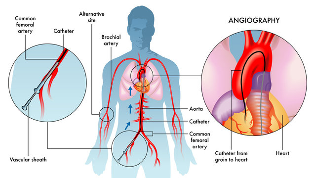
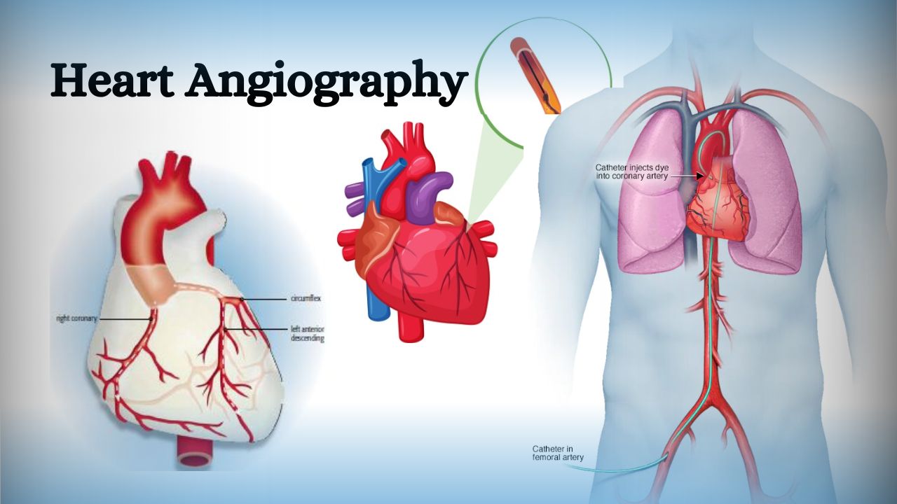
- What Happens During the Angiography?
- Types of Coronary Angiography
- How to Prepare
- Benefits of a Coronary Angiography
- Evaluate Symptoms: To investigate symptoms such as chest pain (angina), shortness of breath, or other signs of heart disease.
- Assess Heart Function: To evaluate heart function and determine the impact of blockages on blood flow to the heart muscle.
- Plan Treatments: To guide decisions for further treatments, such as angioplasty, stent placement, or coronary artery bypass surgery.
- Post-Heart Attack Evaluation: To assess the extent of damage and blockages after a heart attack.
- Preparation:
- Patient fasts for several hours before the procedure.
- Medical history review and pre-procedure tests (e.g., blood tests, ECG) are conducted.
- An intravenous (IV) line is inserted for administering medications and fluids.
- Local anesthesia is applied to numb the insertion site (usually the groin, wrist, or arm).
- Catheter Insertion:
- A small incision is made at the insertion site.
- A sheath (a short, hollow tube) is placed into the artery.
- A catheter (a thin, flexible tube) is inserted through the sheath and guided through the blood vessels to the coronary arteries.
- Contrast Dye Injection:
- A contrast dye is injected through the catheter into the coronary arteries.
- The dye helps to outline the arteries on X-ray images.
- Imaging:
- X-ray images (angiograms) are taken as the dye flows through the coronary arteries.
- The images reveal any blockages, narrowing, or abnormalities in the arteries.
- Invasive Coronary Angiography (ICA):
- Traditional method using a catheter inserted through an artery in the groin, wrist, or arm.
- Involves the injection of contrast dye and X-ray imaging.
- Computed Tomography Coronary Angiography (CTCA):
- Non-invasive method using computed tomography (CT) scanning technology.
- Involves the injection of contrast dye through a vein in the arm.
- Produces high-resolution 3D images of the coronary arteries.
- Magnetic Resonance Coronary Angiography (MRCA):
- Non-invasive method using magnetic resonance imaging (MRI) technology.
- Does not always require contrast dye, but gadolinium-based contrast agents may be used for better visualization.
- Medical Consultation
- Medication Adjustments
Fasting
- Pre-Procedure Tests
- Arrange Transportation
- Review Procedure Details
- Accurate Diagnosis
- Guides Treatment Decisions
- Immediate Treatment
- Assessment of Heart Function
- Prevention of Heart Attacks
